3D Model
Article
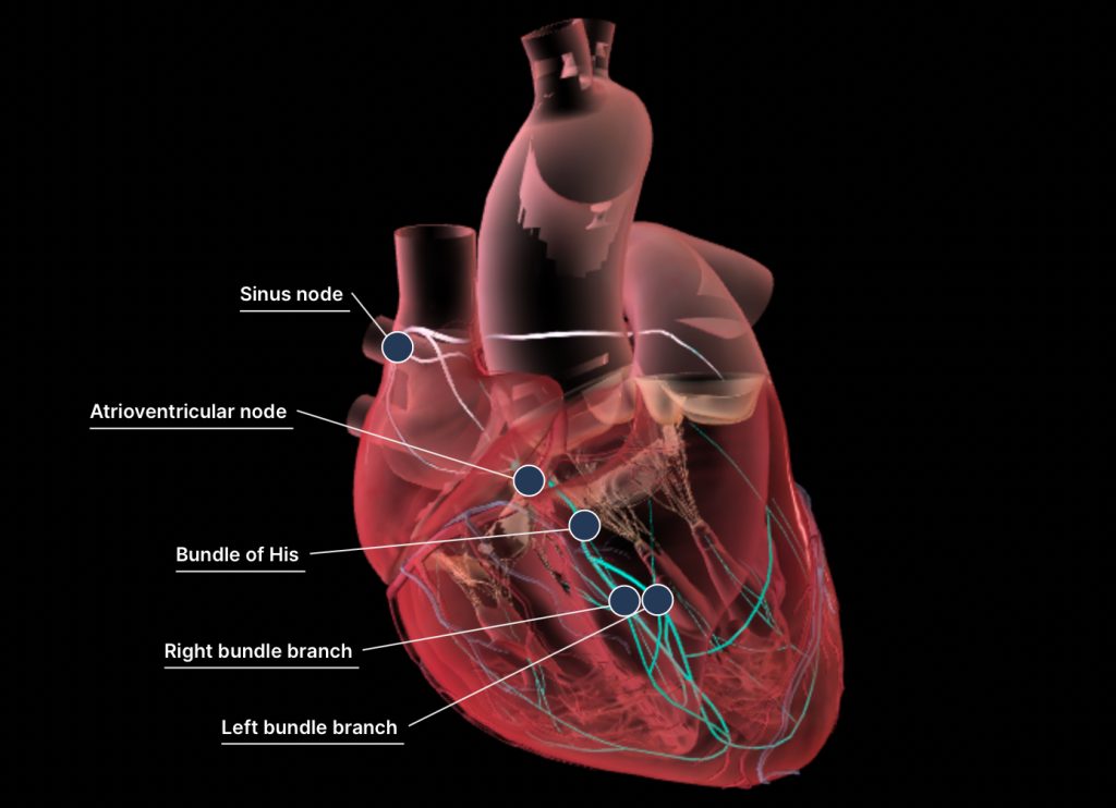
1. SA Node (Sinoatrial Node): Initiates the electrical impulse, causing atrial depolarization (P wave) and atrial contraction.
2. Inter-nodal Bundles: Transmit the electrical signal from the SA node to the AV node.
3. AV Node (Atrioventricular Node): Delays the signal slightly to allow the atria to fully contract before ventricular contraction begins.
4. AV Bundle (Bundle of His): Carries the impulse from the AV node to the ventricles.
5. Purkinje Fibers: Distribute the impulse throughout the ventricles, leading to ventricular depolarization (QRS complex) and contraction.
6. Ventricular Repolarization (T Wave): Relaxation of the ventricles, completing the cycle.
These events ensure the heart beats in a coordinated manner, allowing efficient blood flow.
https://en.m.wikipedia.org/wiki/Cardiac_cycle
Lab — Blood Pressure
Introduction
The webpage focuses on the cardiovascular system, covering blood components (red/white blood cells, platelets, plasma) and their functions, blood vessel pathways, and the structure and function of the heart. It provides details on blood composition, hematocrit levels, and types of plasma proteins. Additionally, it explains blood’s oxygenation states and circulatory routes. For further details, you can read it here:
Videos
Methods of measuring blood pressure:
Process and what you can learn form the lab: